色标引导长脉宽532/1064nm KTP/Nd:YAG激光治疗痤疮后红斑
2026年10月6日24 min read
Journal of Cosmetic Dermatology
结论与实践要点
临床定位与机制: 痤疮后红斑源于炎症后血管扩张与血管重塑。532 nm KTP 被氧合血红蛋白强吸收,适合浅表粉红/红斑血管;1064 nm Nd:YAG 穿透更深,用于紫红/较深血管。本文"色标引导"即按皮损颜色匹配波长(粉红/红→532 nm,紫红→1064 nm,二者兼具则双波长),以提升靶向性并减少周围正常皮肤热损伤。
操作要点: 先以全脸低能量 532 nm KTP 扫描均匀退红,再按颜色定点加强;3 次、间隔 4 周。疼痛轻、停工期短,适合门诊常规开展,本研究 35.29% 患者合并轻度活动期痤疮亦纳入且未出现相关恶化。可将该方案作为 PAE 的常规光电选择之一。
风险与局限: 疗效证据来自单中心回顾性无对照队列,自发缓解不能完全排除,应设定合理预期并告知需多次治疗。对伴明显毛细血管扩张或酒渣鼻样红斑者,可考虑联合其他血管靶向手段(如脉冲染料激光)。随访显示 6 个月内疗效稳定,但长期(>6 个月)维持数据仍缺,需个体化复诊评估。
PurposePost‐acne erythema (PAE) is a common sequela of inflammatory acne. Pulsed dye laser is effective for vascular erythema but is associated with purpura and downtime. This study evaluated the real‐world effectiveness and tolerability of a standardized, color‐guided long‐pulsed 532/1064‐nm KTP/Nd:YAG protocol for PAE.
MethodsThis single‐center retrospective cohort study screened 147 patients and included 136 eligible patients. All patients completed three sessions at 4‐week intervals. Each session comprised low‐fluence full‐face 532‐nm KTP scanning followed by lesion‐targeted treatment: 532‐nm KTP for pink‐to‐red lesions and 1064‐nm Nd:YAG for violaceous lesions, with both wavelengths used when both color patterns were present. Outcomes included an adapted CAT score, erythematous area percentage, Investigator Global Assessment, satisfaction, pain, 6‐month quantitative stability, clinical worsening, and adverse events.
ResultsThe median CAT score decreased from 5 to 1, and the median erythematous area percentage decreased from 4.50% to 1.00% (both p < 0.001). Median CAT improvement rates were 58.34%, 84.52%, and 87.50% in low‐, intermediate‐, and high‐severity groups, respectively; the between‐group difference was not significant ( p = 0.10). All patients in this cohort achieved at least 25% improvement on Investigator Global Assessment. Satisfaction was 97.79%, and the median pain score was 2/10. Transient erythema resolved within 1–3 days. One patient (0.74%) developed purpura with a small blister; no pigmentary sequelae or scarring occurred. At 6 months, CAT score and erythematous area percentage remained comparable with the 1‐month posttreatment values in the overall cohort ( p = 0.285 and p = 0.346, respectively) and across baseline severity subgroups (all p > 0.05).
ConclusionThese findings suggest that a standardized three‐session, color‐guided long‐pulsed 532/1064‐nm KTP/Nd:YAG protocol may be an effective and well‐tolerated option for PAE, with exploratory 6‐month quantitative stability, warranting confirmation in prospective controlled studies.
Keywords: dual‐wavelength laser、KTP laser、Nd:YAG laser、post‐acne erythema、real‐world evidence、vascular laser
1
Post‐acne erythema (PAE) is a common and often persistent sequela of inflammatory acne vulgaris. It presents as pink‐to‐violaceous macules or patches at sites of previously inflamed lesions and may persist for months after active inflammation resolves [ 1 ]. Although nonscarring, PAE can cause substantial cosmetic concern and psychosocial distress. It frequently coexists with postinflammatory hyperpigmentation and acne scarring, further impairing quality of life and treatment satisfaction.
Persistent vasodilation, vascular remodeling, and residual inflammation contribute to PAE. Topical brimonidine and tranexamic acid may transiently reduce erythema through vascular modulation, but their effects are often limited [ 2 ]. Vascular‐targeted laser and light‐based therapies are therefore important treatment options. Pulsed dye laser (PDL), potassium titanyl phosphate (KTP) laser, neodymium‐doped yttrium aluminum garnet (Nd:YAG) laser, and intense pulsed light have been investigated for acne‐associated erythema. The 595‐nm PDL has demonstrated favorable efficacy [ 3 ], but purpura, transient pigmentary change, and downtime may reduce acceptability. Its penetration may also be insufficient for lesions with deeper vascular components.
Wavelength selection is guided by selective photothermolysis, which considers hemoglobin absorption, tissue penetration, and epidermal melanin exposure. The 532‐nm KTP wavelength is strongly absorbed by oxyhemoglobin and preferentially targets superficial dermal vessels, making it suitable for pink‐to‐red lesions [ 4 , 5 , 6 , 7 ]. In contrast, the 1064‐nm Nd:YAG laser penetrates more deeply and has lower melanin absorption, enabling treatment of deeper vascular structures with reduced epidermal exposure [ 8 , 9 ]. These complementary properties support color‐guided wavelength selection for heterogeneous PAE lesions.
Clinical studies support the 532‐nm KTP laser for superficial facial vascular lesions and the 1064‐nm Nd:YAG laser for deeper vascular targets [ 5 , 6 , 7 , 8 , 9 , 10 ]. Additional evidence on dye laser use for vascular lesions and erythematous scars further supports tailored laser protocols in cosmetic dermatology [ 11 ]. However, evidence for a standardized protocol combining full‐face 532‐nm scanning with lesion‐level wavelength selection according to erythema color remains limited in PAE. Its effectiveness, tolerability, and posttreatment stability in routine practice are also insufficiently characterized. Therefore, this retrospective real‐world study evaluated a three‐session, color‐guided long‐pulsed 532/1064‐nm KTP/Nd:YAG protocol in 136 patients with PAE, assessing effectiveness, safety, patient satisfaction, and 6‐month quantitative stability.
2
2.1
This single‐center retrospective cohort study was conducted at the Department of Dermatology, Jiangsu Provincial Geriatric Hospital. Medical records of patients with PAE treated with a long‐pulsed 532/1064‐nm KTP/Nd:YAG laser between October 2024 and October 2025 were reviewed.
2.2
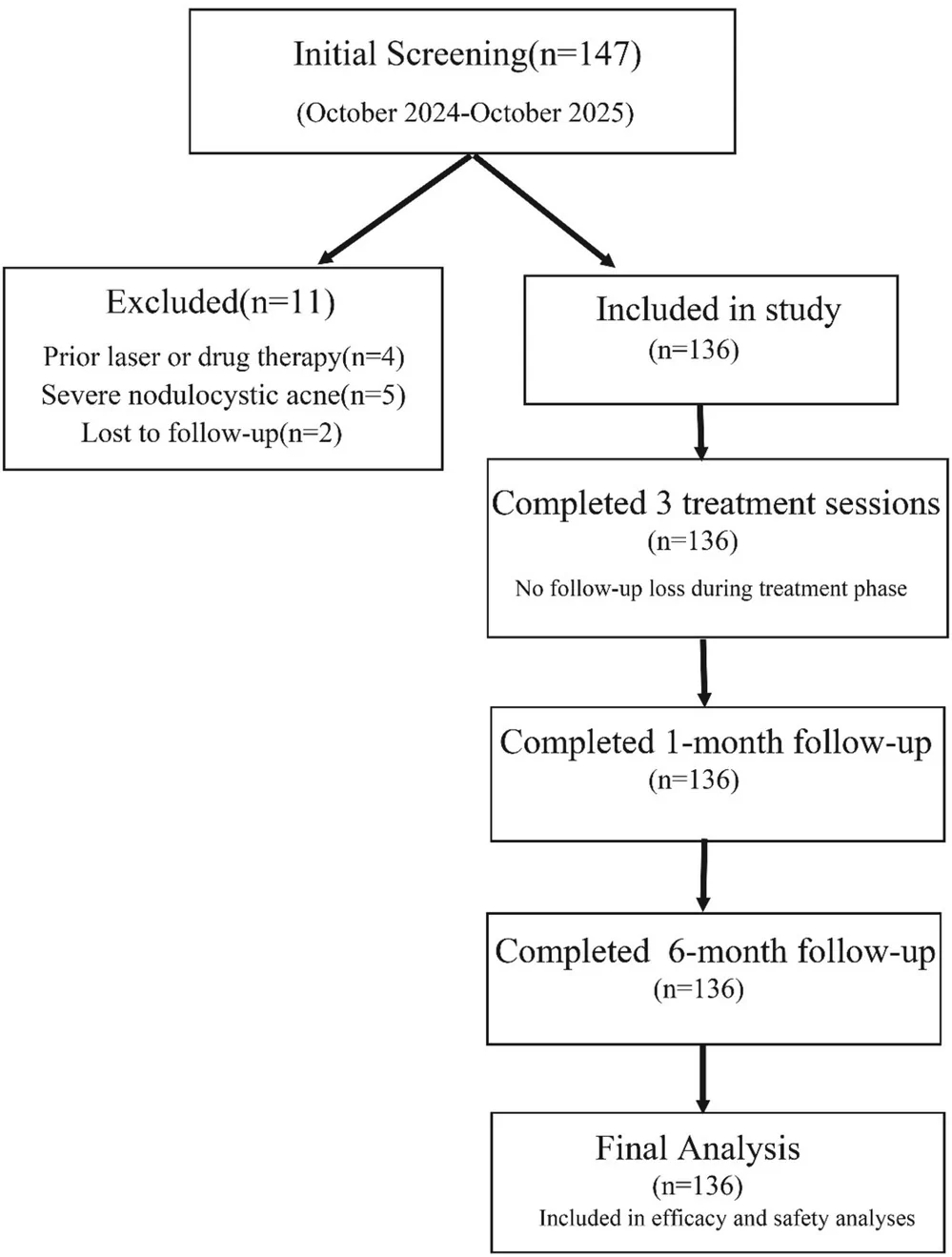
Eligible patients were aged ≥ 18 years, had clinically diagnosed PAE of ≥ 3 months' duration, completed three treatment sessions at 4‐week intervals, and attended follow‐up at 1 and 6 months after the final session. Exclusion criteria were incomplete key follow‐up data; laser, intense pulsed light, systemic, or topical treatment likely to affect erythema within 3 months before treatment; other energy‐based procedures during the study period; severe nodulocystic acne; active cutaneous infection; keloid tendency; or concomitant dermatoses interfering with efficacy assessment. Patient selection is shown in Figure 1 .

FIGURE 1 Patient selection flowchart. Of 147 patients screened, 11 were excluded for prior laser or drug therapy (n = 4), severe nodulocystic acne (n = 5), or loss to follow‐up (n = 2). A total of 136 patients were included in the complete‐case analysis, completed three treatment sessions, and attended both the 1‐ and 6‐month follow‐up assessments.2.3
Treatments were performed using a long‐pulsed 532/1064‐nm laser system (Excel V; Cutera Inc., Brisbane, CA, USA). All patients received three sessions at 4‐week intervals, and each session followed the same two‐step treatment algorithm: standardized low‐fluence full‐face 532‐nm KTP scanning followed by color‐guided lesion‐targeted treatment.
First, the entire face was scanned with the 532‐nm KTP laser using a 10‐mm spot size, pulse duration of 10–15 ms, fluence of 3.0–4.0 J/cm 2 , repetition rate of 2 Hz, and two passes. This standardized low‐fluence step addressed diffuse erythema and concomitant mild inflammatory activity.
Second, lesion‐targeted treatment was selected according to erythema color. Pink‐to‐red lesions were treated with the 532‐nm KTP laser using a 5–7‐mm spot size, pulse duration of 10–12 ms, and fluence of 6.0–8.0 J/cm 2 . Violaceous lesions, considered clinically suggestive of relatively deeper vascular involvement, were treated with the 1064‐nm Nd:YAG laser using a 5–7‐mm spot size, pulse duration of 30–50 ms, and fluence of 50–70 J/cm 2 . Patients with both color patterns received both targeted wavelengths during the same session.
Lesion color was assessed using standardized photographs and a skin imaging system (View Skin, version 1.0.0.0; Hebei Gu'an Jingde Medical Equipment Co., Langfang, Hebei, China) by two dermatologists; a third resolved disagreements. Clinical color served as a pragmatic surrogate for presumed vascular depth. Sapphire‐window contact cooling was maintained at 5°C, and the endpoint was uniform lesion darkening without overt purpura. The same wavelength‐selection algorithm was applied at all three sessions.
2.4
Standardized photographs were obtained before each treatment and at 1 and 6 months after the final session. The prespecified primary efficacy assessment was 1 month after the third treatment.
The primary outcome was an adapted CAT (Clinician Erythema Assessment, Area, and Telangiectasia) score [ 12 ]. Given the absence of a validated PAE‐specific composite score, we adapted the CAT framework to capture clinically relevant dimensions (erythema intensity, area, and telangiectasia) in a single measure, and interpreted it alongside an objective imaging metric (erythematous area percentage). CAT was calculated as the Clinician Erythema Assessment (CEA) score multiplied by the facial area score, plus the telangiectasia score. CEA was graded as 0 = clear, 1 = almost clear, 2 = mild, 3 = moderate, and 4 = severe [ 13 ]. Facial involvement was estimated using the palm method: 1 = one palm area, 2 = two palm areas, and 3 = three palm areas. Telangiectasia was graded as 0 = clear, 1 = focal or mild, and 2 = diffuse or severe. CAT improvement was calculated as (pretreatment CAT—posttreatment CAT)/pretreatment CAT × 100%. Because CAT has not been formally validated for PAE, it was interpreted with the objective erythematous area percentage, defined as the proportion of total facial surface identified as erythematous by the View Skin system [ 14 ].
Secondary outcomes included Investigator Global Assessment (IGA), patient satisfaction, 6‐month quantitative stability, clinical worsening, and safety. IGA was categorized as slight or no improvement (< 25%), moderate improvement (25%–50%), marked improvement (51%–75%), or excellent improvement (>75%) [ 15 ]. Satisfaction was scored as 4 = highly satisfied, 3 = very satisfied, 2 = satisfied, 1 = neutral, and 0 = not satisfied; overall satisfaction was the proportion of patients with scores ≥ 2 [ 15 ]. Clinical worsening was defined as an increase in CAT score or erythematous area percentage from the 1‐month assessment, confirmed by at least two independent investigators. Quantitative 6‐month stability was assessed by comparing CAT score and erythematous area percentage at 6 months with the 1‐month posttreatment values in the overall cohort and baseline severity subgroups.
For the subgroup analyses, patients were categorized into low‐severity (CEA 1), intermediate‐severity (CEA 2), and high‐severity (CEA 3–4) groups.
2.5
Pain during treatment was evaluated using a 10‐point visual analog scale (VAS). Adverse events were recorded, including transient erythema, persistent erythema, edema, purpura, postinflammatory hyperpigmentation, blistering, crusting, and scarring.
2.6
Three dermatologists independently evaluated coded photographs while blinded to treatment parameters and other clinical data. The median of the three ratings was analyzed. Interobserver reliability of individual ratings was assessed using a two‐way mixed‐effects, absolute‐agreement, single‐measure intraclass correlation coefficient (ICC) with 95% confidence intervals.
Statistical analyses were performed using SPSS version 25.0 (IBM Corp., Armonk, NY, USA). Continuous variables are presented as medians with interquartile ranges and categorical variables as counts and percentages. Paired data were compared using the Wilcoxon signed‐rank test. Six‐month CAT and erythematous area assessments were compared with the 1‐month posttreatment values using the Wilcoxon signed‐rank test; these stability analyses were exploratory. Between‐group differences, including ordinal IGA category distributions, were assessed using the Kruskal–Wallis test, with Dunn–Bonferroni post hoc comparisons when appropriate. Other categorical variables were compared using chi‐square or Fisher exact tests. Complete‐case analysis was performed without imputation. No a priori sample‐size calculation was undertaken; all eligible patients treated during the study period were included. All tests were two‐sided, with p < 0.05 indicating statistical significance.
3
3.1
Of 147 patients screened, 11 were excluded for prior laser or drug therapy ( n = 4), severe nodulocystic acne ( n = 5), or loss to follow‐up ( n = 2), and 136 were included in the complete‐case analysis (Figure 1 ). All included patients completed three treatment sessions and the 1‐ and 6‐month follow‐up assessments. Among them, 132 (97.06%) were female and 4 (2.94%) were male. The median age was 25 years (range, 18–45 years). Fitzpatrick skin types II–III accounted for 72.79% of the cohort, whereas types IV and V accounted for 19.85% and 7.35%, respectively. At baseline, 38 patients (27.94%) had low‐severity PAE, 52 (38.24%) had intermediate‐severity PAE, and 46 (33.82%) had high‐severity PAE. Mild active acne and postinflammatory hyperpigmentation were present in 35.29% and 27.21%, respectively. Detailed characteristics are presented in Table 1 .
TABLE 1 Baseline characteristics of included patients (N = 136).
| Characteristic | Value |
|---|---|
| Age, years, median (range) | 25 (18–45) |
| Sex, n (%) | |
| Male | 4 (2.94) |
| Female | 132 (97.06) |
| Fitzpatrick skin type, n (%) | |
| II–III | 99 (72.79) |
| IV | 27 (19.85) |
| V | 10 (7.35) |
| Treatment sessions per patient, n | 3 (all patients) |
| Baseline severity group, n (%) | |
| Low‐severity (CEA 1) | 38 (27.94) |
| Intermediate‐severity (CEA 2) | 52 (38.24) |
| High‐severity (CEA 3–4) | 46 (33.82) |
| Mild active acne, n (%) | 48 (35.29) |
| Postinflammatory hyperpigmentation, n (%) | 37 (27.21) |
Interobserver reliability was excellent, with a single‐measure ICC of 0.93 (95% CI, 0.91–0.95).
3.2
At 1 month after the third treatment, the median CAT score decreased significantly from 5 (interquartile range [IQR], 3–8) at baseline to 1 (IQR, 0–1; p < 0.001). The median CAT improvement rate was 85.71% (IQR, 71.76%–100.00%).
In patients with low‐severity PAE, the median CAT score decreased from 3 (IQR, 2–3) to 1 (IQR, 0–1.25), with a median improvement rate of 58.34% (IQR, 50.00%–100.00%). In the intermediate‐severity group, the median CAT score decreased from 5 (IQR, 5–5.75) to 1 (IQR, 0–1), corresponding to an improvement rate of 84.52% (IQR, 80.00%–100.00%). In the high‐severity group, the median CAT score decreased from 10 (IQR, 8–13.25) to 1 (IQR, 0.75–2), with an improvement rate of 87.50% (IQR, 78.16%–94.65%). CAT improvement rates did not differ significantly among baseline severity groups ( p = 0.10) (Table 2 and Figure 2 ).
TABLE 2 Changes in CAT score and erythematous area percentage after treatment according to baseline severity.
| Outcome | Baseline | Posttreatment | Improvement rate (%) | Paired p |
|---|---|---|---|---|
| CAT score | ||||
| Overall | 5 (3–8) | 1 (0–1) | 85.71 (71.76–100.00) | < 0.001 |
| Low‐severity | 3 (2–3) | 1 (0–1.25) | 58.34 (50.00–100.00) | < 0.001 |
| Intermediate‐severity | 5 (5–5.75) | 1 (0–1) | 84.52 (80.00–100.00) | < 0.001 |
| High‐severity | 10 (8–13.25) | 1 (0.75–2) | 87.50 (78.16–94.65) | < 0.001 |
| Erythematous area percentage (%) | ||||
| Overall | 4.50 (2.00–9.00) | 1.00 (1.00–2.00) | 66.67 (50.00–76.10) | < 0.001 |
| Low‐severity | 4.00 (2.00–6.00) | 1.00 (1.00–2.00) | 57.14 (50.00–71.25) | < 0.001 |
| Intermediate‐severity | 4.50 (3.00–8.75) | 1.00 (1.00–2.00) | 66.67 (50.00–79.23) | < 0.001 |
| High‐severity | 6.00 (3.00–12.25) | 1.00 (1.00–2.25) | 66.67 (53.63–80.19) | < 0.001 |
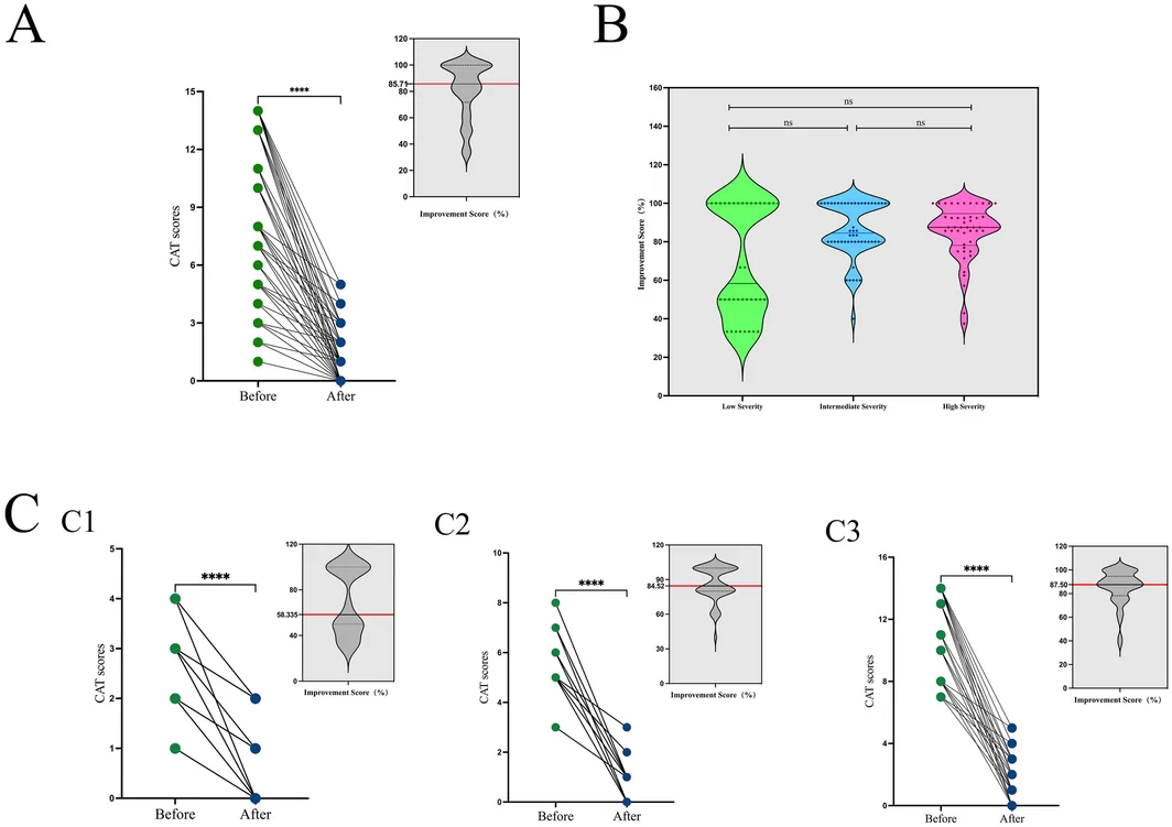
FIGURE 2 (A) CAT scores at baseline and 1 month after the third long‐pulsed KTP/Nd:YAG treatment for PAE. The median CAT score decreased from 5 (IQR, 3–8) to 1 (IQR, 0–1; p < 0.001). (B) Median CAT improvement rates in low‐, intermediate‐, and high‐severity groups were 58.34%, 84.52%, and 87.50%, respectively; the between‐group difference was not statistically significant (Kruskal–Wallis test, p = 0.10). (C) CAT scores before and after treatment according to baseline severity: (C1) low‐severity group (CEA 1, n = 38), from 3 (IQR, 2–3) to 1 (IQR, 0–1.25); (C2) intermediate‐severity group (CEA 2, n = 52), from 5 (IQR, 5–5.75) to 1 (IQR, 0–1); and (C3) high‐severity group (CEA 3–4, n = 46), from 10 (IQR, 8–13.25) to 1 (IQR, 0.75–2). All within‐group differences were significant (p < 0.001). Statistical annotations: **** indicates p < 0.0001, and ns indicates a nonsignificant comparison. In the inset violin plots in panels A and C1‐C3, the red horizontal line marks the median CAT improvement rate.3.3
The median erythematous area percentage decreased significantly from 4.50% (IQR, 2.00%–9.00%) at baseline to 1.00% (IQR, 1.00%–2.00%) at 1 month after the third treatment ( p < 0.001). The median improvement rate for the overall cohort was 66.67% (IQR, 50.00%–76.10%).
Median erythematous area improvement rates were 57.14% (IQR, 50.00%–71.25%), 66.67% (IQR, 50.00%–79.23%), and 66.67% (IQR, 53.63%–80.19%) in the low‐, intermediate‐, and high‐severity groups, respectively. The overall difference among groups was statistically significant ( p < 0.001). Dunn–Bonferroni post hoc analysis identified a significant difference between the low‐ and high‐severity groups (adjusted p < 0.05), whereas the low‐ versus intermediate‐severity and intermediate‐ versus high‐severity comparisons were not significant (Table 2 ).
3.4
At 1 month after the third treatment, all 136 patients achieved at least moderate improvement (≥ 25%) on IGA. The ordinal distribution of IGA categories did not differ significantly among baseline severity groups ( p = 0.345; Table 3 ). Representative clinical photographs are shown in Figure 3 .
TABLE 3 Investigator Global Assessment at 1 month after the third treatment according to baseline severity.
| Baseline severity | < 25% | 25%–50% | 51%–75% | > 75% | p |
|---|---|---|---|---|---|
| Low‐severity ( n = 38) | 0 (0.0) | 11 (28.9) | 9 (23.7) | 18 (47.4) | 0.345 |
| Intermediate‐severity ( n = 52) | 0 (0.0) | 9 (17.3) | 17 (32.7) | 26 (50.0) | |
| High‐severity ( n = 46) | 0 (0.0) | 6 (13.0) | 13 (28.3) | 27 (58.7) |
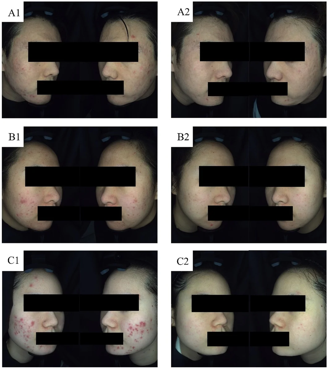
FIGURE 3 Representative standardized clinical photographs obtained at baseline and 1 month after the third long‐pulsed KTP/Nd:YAG treatment. (A) Low‐severity PAE before treatment (A1) and after three sessions (A2). (B) Intermediate‐severity PAE before treatment (B1) and after three sessions (B2). (C) High‐severity PAE before treatment (C1) and after three sessions (C2).3.5
The median pain visual analog scale score was 2 (IQR, 1–3). Transient erythema was observed in 121 patients (88.97%) and resolved spontaneously within 1–3 days. One patient (0.74%) developed immediate purpura followed by a small blister 12 h later; both resolved without treatment‐emergent postinflammatory hyperpigmentation or scarring. No persistent erythema, edema, infection, crusting, or scarring was observed.
At the 6‐month follow‐up, 133 patients (97.79%) reported satisfaction scores ≥ 2, and 3 (2.21%) rated the outcome as neutral. Quantitative follow‐up showed no significant change from 1 month to 6 months in CAT score [1 (0–1) vs. 1 (0–1), p = 0.285] or erythematous area percentage [1.00% (1.00–2.00) vs. 1.00% (1.00–2.00), p = 0.346] in the overall cohort. Similar findings were observed across low‐, intermediate‐, and high‐severity groups for CAT score ( p = 0.206, 0.564, and 0.317, respectively) and erythematous area percentage ( p = 0.102, 0.480, and 0.317, respectively) (Table 4 ).
TABLE 4 Six‐month stability of CAT score and erythematous area percentage.
| Outcome | 1 month | 6 months | p |
|---|---|---|---|
| CAT score | |||
| Overall | 1 (0–1) | 1 (0–1) | 0.285 |
| Low‐severity | 1 (0–1.25) | 1 (0–2) | 0.206 |
| Intermediate‐severity | 1 (0–1) | 1 (0–1) | 0.564 |
| High‐severity | 1 (0.75–2) | 1 (0.75–2) | 0.317 |
| Erythematous area percentage (%) | |||
| Overall | 1.00 (1.00–2.00) | 1.00 (1.00–2.00) | 0.346 |
| Low‐severity | 1.00 (1.00–2.00) | 1.00 (1.00–2.00) | 0.102 |
| Intermediate‐severity | 1.00 (1.00–2.00) | 1.00 (1.00–2.00) | 0.480 |
| High‐severity | 1.00 (1.00–2.25) | 1.00 (1.00–3.00) | 0.317 |
4
This single‐center retrospective real‐world study of 136 patients with PAE found that a standardized three‐session, color‐guided long‐pulsed 532‐nm KTP/1064‐nm Nd:YAG protocol was associated with significant improvement in clinician‐assessed and instrument‐based outcomes, low procedural pain, limited downtime, and high patient satisfaction. The cohort included varied baseline severities, Fitzpatrick phototypes, and concomitant mild acne, reflecting routine clinical heterogeneity. Treatment exposure was uniform, with all patients completing three sessions at 4‐week intervals. Although the uncontrolled design precludes causal inference and spontaneous improvement cannot be excluded, concordant changes across complementary outcomes support the protocol's clinical feasibility. Exploratory quantitative follow‐up further showed that CAT score and erythematous area percentage remained statistically comparable between the 1‐ and 6‐month assessments in the overall cohort and baseline severity subgroups.
Persistent vasodilation and vascular remodeling after inflammation contribute to PAE [ 16 ], supporting vascular‐targeted treatment. The 532‐nm KTP wavelength is strongly absorbed by oxyhemoglobin and is suited to superficial dilated vessels [ 4 , 5 , 6 , 7 ]. KTP treatment may provide improvement comparable to PDL for superficial facial vascular lesions, although prolonged posttreatment erythema has been reported [ 5 ]. In our cohort, conservative fluence, longer pulse durations, a nonpurpuric endpoint, and sapphire‐window contact cooling were associated with a median pain score of 2/10. Posttreatment erythema resolved within 1–3 days, and no persistent edema occurred, indicating limited recovery time.
The 1064‐nm Nd:YAG laser provides greater dermal penetration and lower epidermal melanin absorption than 532‐nm treatment [ 8 , 17 , 18 , 19 ]. These properties may benefit violaceous lesions with presumed deeper vascular involvement and patients with darker phototypes. Previous studies reported erythema improvement comparable to 595‐nm PDL, with fewer adverse events [ 9 ]. In our cohort, 27.21% of patients had Fitzpatrick skin types IV–V, and no treatment‐emergent persistent postinflammatory hyperpigmentation occurred. This finding is consistent with the reported tolerability of long‐pulsed 1064‐nm treatment [ 20 ]. However, safety was not analyzed separately by phototype or targeted wavelength, precluding comparative conclusions.
The protocol was defined by a standardized two‐step structure. All patients received two low‐fluence full‐face 532‐nm scanning passes, followed by color‐guided lesion treatment: 532 nm for pink‐to‐red lesions and 1064 nm for violaceous lesions. Patients with mixed color patterns received both targeted wavelengths during the same session. This approach is clinically relevant because PAE may contain heterogeneous vascular patterns within the same face, making a fixed single‐wavelength protocol less adaptable to routine practice. The full‐face passes were intended to address diffuse erythema and concomitant mild inflammatory activity [ 21 , 22 , 23 ], although acne‐specific outcomes were not assessed. Vascular depth was estimated from clinical color rather than confirmed by vascular imaging. Wavelength‐specific exposures and outcomes were not analyzed separately. Therefore, the relative contributions of the full‐face and targeted components cannot be distinguished.
CAT improvement did not differ significantly among baseline severity groups, whereas erythematous area improvement differed between the low‐ and high‐severity groups. This discrepancy may reflect the dimensions captured by each measure. CAT integrates erythema intensity, lesion extent, and telangiectatic change, whereas erythematous area percentage quantifies only the proportion of facial surface classified as erythematous. Floor effects in low‐severity disease may also have limited measurable relative change. Because CAT was developed for erythematotelangiectatic rosacea rather than PAE, its use should be considered an adapted multidimensional assessment and interpreted with the objective imaging outcome.
Transient erythema was the most frequent adverse event and resolved within several days. Only one patient developed purpura followed by a small blister; both resolved without pigmentary sequelae or scarring. Previous PDL studies in PAE or erythematous acne scars reported higher purpura rates [ 24 ]. Such indirect comparisons require caution because patient characteristics, treatment parameters, endpoints, and adverse‐event definitions differ across studies. The low purpura rate may relate to conservative fluence, longer pulse durations, a nonpurpuric endpoint, and contact cooling [ 10 , 25 , 26 ], but these explanations cannot be confirmed without a comparative design. The study's real‐world value lies in showing that a prespecified color‐guided algorithm can be implemented across a heterogeneous cohort with low procedural discomfort and limited downtime.
Several limitations should be acknowledged. First, the absence of an untreated, single‐wavelength, or PDL comparator prevents causal attribution and direct efficacy or safety comparisons. Second, the retrospective single‐center design and exclusion of patients with incomplete follow‐up may have introduced selection and attrition bias; the predominance of women further limits generalizability. Third, clinical color was a pragmatic surrogate for vascular depth, and wavelength‐specific exposures and outcomes were not analyzed separately. Fourth, the adapted CAT score has not been validated for PAE, and active acne may have influenced erythema measurements; no multivariable adjustment was performed for phototype or postinflammatory hyperpigmentation. Finally, no a priori sample‐size calculation was performed, and follow‐up was limited to 6 months. Although CAT and erythematous area were repeated at 6 months, this stability analysis was exploratory and lacked a comparator or longer‐term data. Nevertheless, the uniform schedule, standardized assessments, blinded photographic evaluation, excellent interobserver reliability, objective imaging, and consistent within‐patient changes strengthen the internal coherence of the findings.
5
Overall, this study provides real‐world evidence that a standardized three‐session protocol combining full‐face low‐fluence 532‐nm KTP scanning with color‐guided lesion‐targeted 532‐nm KTP and/or 1064‐nm Nd:YAG treatment was associated with improvement in PAE, low procedural pain, limited downtime, and favorable tolerability. Exploratory quantitative follow‐up showed no statistically significant change in CAT score or erythematous area between 1 and 6 months after treatment. The strategy may support individualized wavelength selection when lesions of different colors coexist; however, its comparative value over single‐wavelength treatment or PDL remains uncertain. Prospective multicenter controlled studies incorporating predefined wavelength‐allocation criteria, objective vascular imaging, validated patient‐reported outcomes, and longer follow‐up are warranted.
Jiayi Jiang and Bingrong Zhou conceived the study, and all authors contributed to its design. Jiayi Jiang and Xiaoqing Wang performed the treatments. Yan Tang and Jingwen Jiang coordinated patient enrollment, questionnaires, and follow‐up. Jiayi Jiang, Ying Yang, and Xiaoqing Wang performed the photographic assessments and statistical analyses. Jiayi Jiang drafted the manuscript; Ying Yang and Bingrong Zhou revised it. All authors approved the final version.
This work was supported by the National Natural Science Foundation of China (grant no. 82073472); the Priority Academic Program Development of Jiangsu Higher Education Institutions; the Nantong Young Medical Expert Training Plan; and the Outstanding Young and Middle‐Aged Talents Support Program of the First Affiliated Hospital of Nanjing Medical University (Jiangsu Province Hospital).
This study was conducted in accordance with the Declaration of Helsinki and approved by the Ethics Committee of Jiangsu Provincial Geriatric Hospital (approval no. 0921‐1). Written informed consent was obtained from all participants.
Written informed consent was obtained from all study participants.
The authors declare no conflicts of interest.
声明:中文精读 · 仅供学术参考。内容来自公开文献检索,不代表本人观点,不构成诊疗建议。 医疗美容需在正规医疗机构由执业医师实施。
基于相同主题推荐 · 继续深挖
单中心回顾性真实世界研究(n=136 痤疮后红斑 PAE 患者):标准化色标引导方案为全脸低能量 532 nm KTP 扫描,再按皮损颜色定点(粉红/红用 532 nm KTP,紫
背景:评估羟基磷灰石钙(CaHA)填充注射联合 CO2 剥脱点阵激光(AFL)对萎缩性痤疮瘢痕的疗效与安全。
设备概述:新一代 Fraxel FTX(Solta Medical,2025)为 1550nm 掺铒玻璃光纤 + 1927nm 铥光纤非剥脱点阵激光,较上一代 Fraxel DUA